Rib Cage Anatomy Posterior View - Human Skeleton System Rib Cage Described With Labels Anatomy Posterior View Stock Illustration Illustration Of Elbow Limbs 176983816 / We did not find results for:
Rib Cage Anatomy Posterior View - Human Skeleton System Rib Cage Described With Labels Anatomy Posterior View Stock Illustration Illustration Of Elbow Limbs 176983816 / We did not find results for:. Check spelling or type a new query. Posterior all the twelve ribs articulate posteriorly with the vertebrae of the spine. From www.meddean.luc.edu alison.com has been visited by 100k+ users in the past month learn the basic anatomy and physiology of the human body with this free online course. There are twelve (12) pairs of ribs and all articulate posteriorly with the thoracic vertebrae. These pass from the inferior edge of the costal groove to the superior margins of the ribs below.
The posterior end of a typical rib is called the head of the rib (see chapter 7.3 figure 7.3.8). 35, 36, 37 the external oblique muscle flap is capable of reaching the third ipsilateral rib space superiorly and up to 5 cm beyond. An mri scan gives the doctor a detailed view of your rib cage and surrounding muscles, organs, and tissue. These pass from the inferior edge of the costal groove to the superior margins of the ribs below. Ribs (ap view) the ribs ap view is a specific projection employed in the assessment of the posterior ribs.
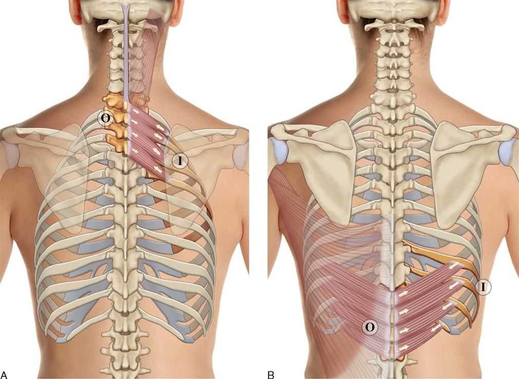
Shutterstock Puzzlepix from image.shutterstock.com Lateral view of a pair of ribs articulating with the thoracic vertebrae. Each rib forms two joints: It is innervated by the first four lumbar nerves, plus the twelfth thoracic nerve. The first 7 pairs use the sternum as their anchor via the rib's individual costal cartilage as an attachment vessel. They articulate at the costochondral joints with some exceptions. The eleven pairs of internal intercostal muscles are found posterior to the external intercostals. These pass from the inferior edge of the costal groove to the superior margins of the ribs below. The serratus posterior inferior originates on the spinous processes of t11 through l2 and inserts on the inferior posterior aspect of ribs 9 through 12.
Rib cage posterior view in humans, the rib cage, also known as the thoracic cage, is a bony and cartilaginous structure which surrounds the thoracic cavity and supports the pectoral girdle (shoulder girdle), forming a core portion of the human skeleton.
These muscle fibres extend in a posteroinferior direction and again pass in an oblique manner. The upper edge is round and the lower sharp. Rib cage posterior view in humans, the rib cage, also known as the thoracic cage, is a bony and cartilaginous structure which surrounds the thoracic cavity and supports the pectoral girdle (shoulder girdle), forming a core portion of the human skeleton. This region articulates primarily with the costal facet located on the body of the same numbered thoracic vertebra and to a lesser degree, with the costal facet located on the body of the next higher vertebra. Arises from the posterior border of the iliac crest and inserts on the first to fourth lumbar vertebrae plus the twelfth rib. They articulate at the costovertebral joints at the head of the rib and at the costotransverse joints with the tubercle. Unlike a standard chest radiograph, this projection applies a lower kv higher mas technique to highlight bony structures. Posterior all the twelve ribs articulate posteriorly with the vertebrae of the spine. Anteriorly, most are attached directly to the sternum. Extent of the region and the articulations with the rib cage. The ribs are attached to corresponding thoracic vertebrae posteriorly. Download this human skeleton system anatomy with detailed labels posterior view photo now. Chest bone, ribs, lung, heart, xiphoid process, sternum anatomy.
There are 24 ribs in the human body, divided into two sets of 12 curved, flat bones. Check spelling or type a new query. At the chest, many rib bones connect to the sternum via costal cartilage,. Unlike a standard chest radiograph, this projection applies a lower kv higher mas technique to highlight bony structures. Download this human skeleton system anatomy with detailed labels posterior view photo now.
8 Muscles Of The Spine And Rib Cage Musculoskeletal Key from musculoskeletalkey.com A rib has a flat body, as you can see from the picture of the anatomy of the human rib cage. 35, 36, 37 the external oblique muscle flap is capable of reaching the third ipsilateral rib space superiorly and up to 5 cm beyond. These muscle fibres extend in a posteroinferior direction and again pass in an oblique manner. The rib cage is a bony structure found in the chest (thoracic cavity). With each succeeding rib, from the first, or uppermost, the curvature of the rib cage becomes more open. In this image, you will find common carotid arteries, internal jugular vein, subclavian artery, subclavian vein, heart, right lung, 6th rib, diaphragm, costal cartilage in it. The posterior end of a typical rib is called the head of the rib (see chapter 7.3 figure 7.3.8). We did not find results for:
A rib has a flat body, as you can see from the picture of the anatomy of the human rib cage.
Chest bone, ribs, lung, heart, xiphoid process, sternum anatomy. In the inferior pair of ribs (i), the posterior rib (arrow) is slightly lower than the anterior rib. The posterior (dorsal) and anterior (ventral) cavities are each subdivided into smaller cavities. The superior fibres originate from the spinous processes of the c7 to t3 vertebrae and attach to the superior borders of ribs two to four. Anteriorly, most are attached directly to the sternum. Of all 24 ribs, the It is made up of 12 pairs of ribs. In the posterior (dorsal) cavity, the cranial cavity houses the brain, and the spinal cavity (or vertebral cavity) encloses the spinal cord. Posterior all the twelve ribs articulate posteriorly with the vertebrae of the spine. Each one is attached by cartilage at the back to the thoracic vertebrae. They articulate at the costochondral joints with some exceptions. The serratus posterior inferior originates on the spinous processes of t11 through l2 and inserts on the inferior posterior aspect of ribs 9 through 12. If you're experiencing chronic pain, your doctor may order a bone scan.
Lateral view of a pair of ribs articulating with the thoracic vertebrae. The serratus posterior inferior originates on the spinous processes of t11 through l2 and inserts on the inferior posterior aspect of ribs 9 through 12. Each are symmetrically paired on a right and left side. A rib has a flat body, as you can see from the picture of the anatomy of the human rib cage. Arises from the posterior border of the iliac crest and inserts on the first to fourth lumbar vertebrae plus the twelfth rib.
3d Illustration Of Human Skeleton System Rib Cage With Labels Anatomy Anterior View Canstock from comps.canstockphoto.com This region articulates primarily with the costal facet located on the body of the same numbered thoracic vertebra and to a lesser degree, with the costal facet located on the body of the next higher vertebra. Just lateral to the tubercle is the angle of the rib, the point at which the rib has its greatest degree of curvature. The first 7 pairs use the sternum as their anchor via the rib's individual costal cartilage as an attachment vessel. Of all 24 ribs, the Related posts of muscle anatomy ribs muscle anatomy posterior. Posterior anatomy of rib cage. The remainder of the rib is the body of the rib (shaft). These pass from the inferior edge of the costal groove to the superior margins of the ribs below.
Ribs (ap view) the ribs ap view is a specific projection employed in the assessment of the posterior ribs.
The human rib cage is made up of 12 paired rib bones; Just lateral to the tubercle is the angle of the rib, the point at which the rib has its greatest degree of curvature. In the anatomical position, the angles align with the medial border of the scapula. They articulate at the costochondral joints with some exceptions. The typical ribs have a generalised structure while the atypical ribs have variations on this structure. Chest bone, ribs, lung, heart, xiphoid process, sternum anatomy. An mri scan gives the doctor a detailed view of your rib cage and surrounding muscles, organs, and tissue. In the inferior pair of ribs (i), the posterior rib (arrow) is slightly lower than the anterior rib. With each succeeding rib, from the first, or uppermost, the curvature of the rib cage becomes more open. The upper edge is round and the lower sharp. There are twelve (12) pairs of ribs and all articulate posteriorly with the thoracic vertebrae. Innervation is from the inferior six intercostal nerves, and blood supply primarily derives from the anterior and collateral branches of the posterior intercostal arteries in the 10th and 11th intercostal spaces. You will also find the xiphoid process, 10th rib, the apex of the heart, the coronary vein of the heart.
Posterior anatomy of rib cage rib cage anatomy. Check spelling or type a new query.


0 Komentar